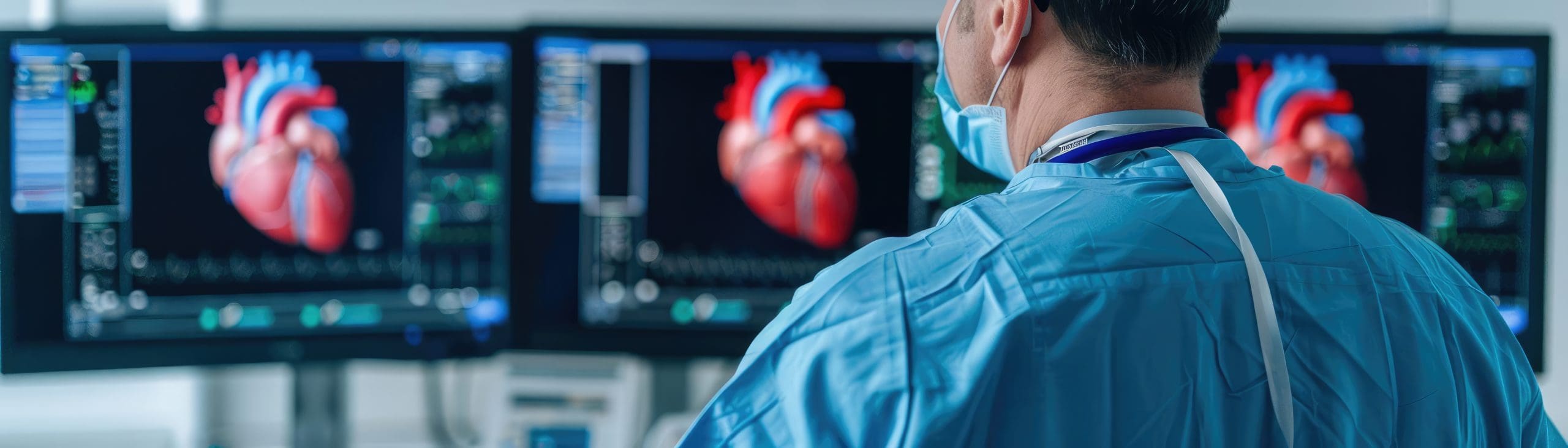
In neurology, timing and clarity are everything. Whether you’re a neurologist evaluating cognitive decline or a radiologist supporting cross-specialty diagnosis, access to detailed, functional brain imaging can significantly influence patient care.
But what happens when that imaging is performed in a portable setting?
Thanks to mobile neurology solutions, PET brain scans can now be performed wherever they’re needed—without sacrificing image quality, interpretation accuracy, or the physician collaboration critical to neurology care.
Here’s a breakdown of how these scans are interpreted and how they fit seamlessly into modern clinical workflows.
Mobile PET systems are designed to deliver high-resolution neuroimaging that matches hospital-based scanners. These systems include:
• PET (Positron Emission Tomography) for functional and metabolic brain imaging using tracers such as FDG
This imaging plays a critical role in detecting early signs of Alzheimer’s disease, frontotemporal dementia, epilepsy, and cerebrovascular disease—conditions frequently evaluated by neurologists and, in some cases, cardiologists.
In mobile neurology, every step—from image acquisition to interpretation—is designed to integrate directly into your existing clinical workflow.
A common question among providers is whether portable imaging results in slower turnaround times or reduced image quality.
The answer is no. Modern Nuclear uses the same imaging protocols, safety standards, and technologist training found in fixed imaging environments.
With a portable unit:
• Board-certified nuclear medicine technologists perform the scan
• Patients undergo PET imaging following appropriate radiotracer uptake
• Advanced image-processing software corrects for anatomical artifacts
• Scans are uploaded to secure cloud-based PACS for rapid, remote interpretation
This ensures clinicians receive diagnostic-quality images with no compromise—ideal for neurologists interpreting neurodegenerative conditions.
Interpretation is a multi-layered process. Although images are acquired on-site, they are read by board-certified radiologists or nuclear medicine physicians, often with specialized expertise in neurodegenerative and cerebrovascular disorders.
Interpreting physicians focus on several key elements:
Brain metabolic patterns linked to neurological disorders
FDG-PET imaging reveals glucose metabolism across specific brain regions, helping differentiate between conditions such as:
• Alzheimer’s disease, which often presents with reduced activity in the posterior parietal and temporal lobes
• Frontotemporal dementia, characterized by decreased uptake in frontal regions
• Parkinson’s disease or Lewy body dementia, which may show distinct patterns including occipital hypometabolism
• Epilepsy, where interictal hypometabolism is frequently localized to the seizure focus
For neurologists, these metabolic patterns help confirm clinical suspicions. For cardiologists managing patients with vascular dementia or mixed pathology, PET imaging can clarify whether neurodegeneration is present or rule it out entirely.
Modern Nuclear also supports integration with quantitative analysis tools that enhance interpretation accuracy, including:
• Normative brain comparisons
• Z-score mapping of regional brain activity
• Pattern-recognition algorithms that support earlier diagnosis
These quantitative insights strengthen diagnostic confidence, particularly in multispecialty cases. Whether managing memory loss or coordinating complex cognitive evaluations, quantitative data adds objective metrics to visual interpretation.
Mobile neurology solutions are particularly well-suited for:
• Memory clinics
• Outpatient neurology practices
• Community hospitals without in-house PET imaging
The workflow is streamlined and efficient:
• The scan is performed on-site at your facility
• Images are uploaded immediately to a secure platform
• Interpreting physicians deliver reports within 24–48 hours
• Reports are tailored to clinical relevance, such as hippocampal hypometabolism for Alzheimer’s disease or parietal-temporal changes linked to vascular decline
The result is faster diagnoses, clearer insights, and stronger collaboration across specialties—without delays or guesswork.
PET imaging is more than a diagnostic test; it’s a decision-making tool that supports early diagnosis, differential evaluation, and treatment planning.
Neurologists can use PET imaging to confirm or rule out diagnoses before initiating costly or invasive therapies. Radiologists can provide fusion reports that align neurological and cardiovascular findings, guiding unified, patient-centered care planning.
Interpreting the Brain from Anywhere—with Accuracy and Speed
Mobile neurology has expanded access to advanced imaging without lowering quality standards. With portable PET imaging units, interpretation remains precise, collaborative, and clinically actionable—regardless of where the scan takes place.
For neurologists managing progressive memory loss or complex neurological presentations, portable PET imaging closes diagnostic gaps and brings clarity to challenging cases.
The brain doesn’t wait—and now, neither do you.
FAQs
Are mobile PET scans suitable for diagnosing dementia or Alzheimer’s disease?
Yes. Portable PET solutions are fully equipped to deliver FDG-PET scans essential for the early detection and differentiation of Alzheimer’s disease and related dementias.
How do I receive the interpreted report from a mobile scan?
Scans are uploaded securely and interpreted remotely by a board-certified radiologist or nuclear medicine physician. Reports are typically delivered within 24–48 hours.
Is image quality comparable to hospital-based scanners?
Yes. Portable PET technology uses high-resolution detector systems and advanced software, ensuring no compromise in image quality or interpretive accuracy.